Un-Numbered equations and images beside lists (Medical Handbooks)
Capture un-numbered equations beside lists at the end of the list item. Capture images beside lists at the end of the list if the image is not cross referenced in the text.
Capture un-numbered equations beside lists at the end of the list item. For images beside lists, if the figures, tables, media, or boxedMatter are not cross referenced in the text then insert the processing instruction at the end of the div2 division that it appears alongside. Set the target for the xref's ref attribute as the div2 element that the figure appears in.
Equations: Manuscript
Equations: XML
<p>
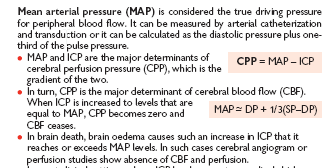
<b>Mean arterial pressure (MAP)</b> is considered the true driving pressure for peripheral
blood flow. It can be measured by arterial catheterization and transduction or it can be
calculated as the diastolic pressure plus one third of the pulse pressure.</p>
<p>
<list class="other"><list1 listType="unstructured">
<item1 id="med-9780198530077-chapter-1-item1-100">
<p>
<enumerator>•</enumerator> MAP and ICP are the major determinants of cerebral
perfusion pressure (CPP), which is the gradient of the two.</p>
<p>
<displayMaths><!-- CPP = MAP – ICP --></displayMaths>
</p>
</item1>
<item1 id="med-9780198530077-chapter-1-item1-101">
<p>
<enumerator>•</enumerator> In turn, CPP is the major determinant of cerebral blood
fl ow (CBF). When ICP is increased to levels that are equal to MAP, CPP becomes zero
and CBF ceases.</p>
<p>
<displayMaths><!--
MAP –~ DP + 1/3(SP–DP) --></displayMaths>
</p>
</item1>
<item1 id="med-9780198530077-chapter-1-item1-102">
<p>
<enumerator>•</enumerator> In brain death, brain oedema causes such an increase in
ICP that it reaches or exceeds MAP levels. In such cases cerebral angiogram or
perfusion studies show absence of CBF and perfusion.</p>
</item1></list1>
</list>
</p>
Images: Manuscript
Images: XML
<p>
<list class="other"><title>
<p>Normal values</p></title><list1 listType="unstructured">
<item1 id="med-9780199512345-chapter-1-item1-1">
<p>
<enumerator>•</enumerator> In general, CVP is 0.67 9 0.4kPa ...</p>
</item1>
<item1 id="med-9780199512345-chapter-1-item1-2">
<p>
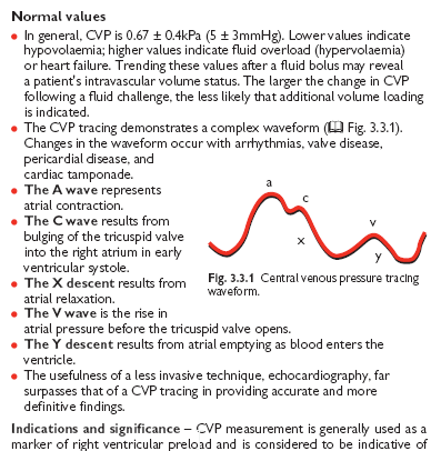
<enumerator>•</enumerator> The CVP tracing demonstrates a complex waveform (<xref ref="med-9780199512345-chapter-1-figureGroup-3.3.1">Fig. 3.3.1</xref>)...</p>
</item1>
<item1 id="med-9780199512345-chapter-1-item1-3">
<p>
<enumerator>•</enumerator> The A wave represents atrial contraction.</p>
</item1>
<item1 id="med-9780199512345-chapter-1-item1-4">
<p>...</p>
</item1>
<item1 id="med-9780199512345-chapter-1-item1-5">
<p>
<enumerator>•</enumerator> The usefulness of a less invasive technique,
echocardiography, far surpasses that of a CVP tracing in providing accurate and more
definitive findings.</p>
</item1></list1>
</list>
</p>
<p><?Insert-Figure ID="med-978019nnnnn-figureGroup-3.3.1"?></p>
<p>
<b>Indications and significance</b> – CVP measurement is generally ...</p>